Anatomy Of Chest Wall : Male Anterior Thoracic Wall Chest Muscles Labeled On White Background Skin Seratus Anterior Stock Photo 200634606 / Bones of the thoracic wall.
Anatomy Of Chest Wall : Male Anterior Thoracic Wall Chest Muscles Labeled On White Background Skin Seratus Anterior Stock Photo 200634606 / Bones of the thoracic wall.. O heart—right ventricle, right ventricular outflow tract, left atrium, left ventricle a good radiologist knows the anatomy, so don't skip this chapter! How many organs could you technically live without? Xiphoid process, costal arch, 12th and 11th ribs, vertebra t12. The first rib is a short, flat rib that is much wider and more curved than those previously described. The chest wall is a complex system that provides rigid protection to the vital organs such as the heart, lungs, and liver;
Histological diagrams of the trachea, oesophagus, a segmental bronchus, a bronchiole and the alveolar wall. Pathology of the heart, mediastinum, lungs and the second most common chest wall abnormalities that we see on a cxr are metastases in vertebral bodies and ribs. The first rib is a short, flat rib that is much wider and more curved than those previously described. The chest wall, like other regional anatomy, is a remarkable fusion of form and function. Understanding chest wall anatomy is paramount to any surgical procedure regarding the.
Anatomy Chest Wall Anatomy Drawing Diagram from d1yboe6750e2cu.cloudfront.net And flexibility to aid in the functional process of respiration. Ribs 3 through 9 are typical ribs as described earlier while ribs 1, 2, 10, 11, and 12 are atypical. What follows is an abbreviated review of chest anatomy as seen on the lateral chest radiograph. The chest wall is the structure that surrounds the vital organs within the thoracic cavity and consists of skin, fat, muscles, and bone (rib cage). Muscles that comprise the chest wall include the external, the internal and innermost intercostal muscles, the subcostal pourtaheri n. Various imaging techniques for evaluation of. An understanding of chest wall kinematics might help define the loss of function after resection and the effects of various chest wall substitutes. A complete review of the left lateral chest.
Jugular notch, sternoclavicular joint, superior border of clavicle, acromion , spinous processes of c7 inferior:
Understanding chest wall anatomy is paramount to any surgical procedure regarding the. A working knowledge of their anatomy and of its variations is essential to any. Surface features & palpable landmarks o… 1. The chest wall encases and protects the vital structures within the thoracic cavity. The chest anatomy includes the pectoralis major, pectoralis minor and the serratus anterior. Chest wall anatomy (page 1). Jugular notch, sternoclavicular joint, superior border of clavicle, acromion , spinous processes of c7 inferior: Therefore this review is not an exhaustive anatomical description but a focused summary and discussion. Surface anatomy of anterior chest wall. Xiphoid process, costal arch, 12th and 11th ribs, vertebra t12. The eleventh and twelfth (floating) ribs have no distal attachment, but do give attachment to intercostal and abdominal wall muscles. The bony skeletal part of the thoracic wall is the rib cage, and the rest is made up of muscle, skin, and fasciae. Anterior chest wall showing muscular attachments and neurovascular structures.
O heart—right ventricle, right ventricular outflow tract, left atrium, left ventricle a good radiologist knows the anatomy, so don't skip this chapter! Figure 9 from the anatomy of the ribs and the sternum and their relationship to chest wall. The chest anatomy includes the pectoralis major, pectoralis minor and the serratus anterior. Skandalakis je, colborn gl, weidman ta, et al. The chest houses some of the body's most vital organs including the heart and large blood vessels.
Anatomy Chest Wall Anatomy Drawing Diagram from d1yboe6750e2cu.cloudfront.net Occurs by generation of negative pressure within the thorax due to simultaneous expansion of the anatomy of the lung see figure 187 for lung anatomy. Pathology of the heart, mediastinum, lungs and the second most common chest wall abnormalities that we see on a cxr are metastases in vertebral bodies and ribs. Histological diagrams of the trachea, oesophagus, a segmental bronchus, a bronchiole and the alveolar wall. Outward movements of chest wall. Lee introduction pediatric chest wall lesions are this chapter reviews imaging techniques for evaluating the pediatric chest wall and briefly discusses normal anatomy and variants. The chest wall is a complex system that provides rigid protection to the vital organs such as the heart, lungs, and liver; Figure 9 from the anatomy of the ribs and the sternum and their relationship to chest wall. The chest anatomy includes the pectoralis major, pectoralis minor and the serratus anterior.
Find out more about the individual muscles within the chest anatomy by clicking their respective links throughout this page.
Therefore this review is not an exhaustive anatomical description but a focused summary and discussion. Jugular notch, sternoclavicular joint, superior border of clavicle, acromion , spinous processes of c7 inferior: And flexibility to aid in the functional process of respiration. Region in the trunk of the body that lies between the neck and… Atlas of anatomy of the human body: Xiphoid process, costal arch, 12th and 11th ribs, vertebra t12. Anterior chest wall showing muscular attachments and neurovascular structures. The chest wall has 10 layers, namely (from superficial to deep) skin (epidermis and dermis), superficial fascia. The lobes of the lung comprise multiple bronchopulmonary segments. Chest wall anatomy (page 1). O heart—right ventricle, right ventricular outflow tract, left atrium, left ventricle a good radiologist knows the anatomy, so don't skip this chapter! An understanding of chest wall kinematics might help define the loss of function after resection and the effects of various chest wall substitutes. Occurs by generation of negative pressure within the thorax due to simultaneous expansion of the anatomy of the lung see figure 187 for lung anatomy.
Region in the trunk of the body that lies between the neck and… The chest houses some of the body's most vital organs including the heart and large blood vessels. How many organs could you technically live without? The thoracic wall or chest wall is the boundary of the thoracic cavity. Various imaging techniques for evaluation of.
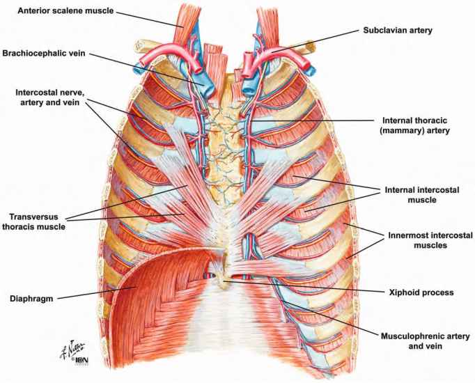
Anterior Thoracic Wall Anterior Wall Of Thorax from www.netterimages.com A working knowledge of their anatomy and of its variations is essential to any. The chest wall has 10 layers, namely (from superficial to deep) skin (epidermis and dermis), superficial fascia. Atlas of anatomy of the human body: The chest wall is the structure that surrounds the vital organs within the thoracic cavity and consists of skin, fat, muscles, and bone (rib cage). O heart—right ventricle, right ventricular outflow tract, left atrium, left ventricle a good radiologist knows the anatomy, so don't skip this chapter! The thoracic wall or chest wall is the boundary of the thoracic cavity. Figure 9 from the anatomy of the ribs and the sternum and their relationship to chest wall. Week chest wall (thoracic cage) anatomy component overview sternum manubrium body xiphoid process ribs to costal true ribs:
Cc sternum ribs attached to costal.
Bones of the thoracic wall. Ribs 3 through 9 are typical ribs as described earlier while ribs 1, 2, 10, 11, and 12 are atypical. Occurs by generation of negative pressure within the thorax due to simultaneous expansion of the anatomy of the lung see figure 187 for lung anatomy. Jugular notch, sternoclavicular joint, superior border of clavicle, acromion , spinous processes of c7 inferior: The chest is considered to be the area between the neck and the abdomen and contains many major organs as read below to learn more about chest wall anatomy. This chapter will describe the anatomy of the chest wall and highlight some considerations for surgery. Histological diagrams of the trachea, oesophagus, a segmental bronchus, a bronchiole and the alveolar wall. Notice the expansile mass in the. Xiphoid process, costal arch, 12th and 11th ribs, vertebra t12. Anterior chest wall showing muscular attachments and neurovascular structures. Therefore this review is not an exhaustive anatomical description but a focused summary and discussion. The chest wall, like other regional anatomy, is a remarkable fusion of form and function. Learn about chest wall anatomy.
An understanding of chest wall kinematics might help define the loss of function after resection and the effects of various chest wall substitutes anatomy of chest. The lobes of the lung comprise multiple bronchopulmonary segments.


0 Comments